WaveQuanta
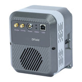
DField-VS-A4 TE-Cooled Spectroscopy CCD, -60 °C Cooling, 30.7 mm Wide Sensor
DField-VS-A4Contact for pricing
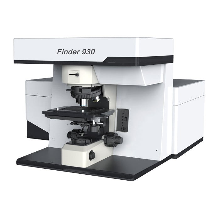
Fully automated laser confocal Raman microscope for Raman and fluorescence mapping in research laboratories.
Finder 930 is a fully automated laser confocal Raman spectroscopy system built around an upright microscope, an image-corrected 320 mm spectrograph and up to three built-in lasers. Laser selection, laser power, confocal pinhole size and optical path correction are motor-controlled from the software. The system software covers Raman mapping, fluorescence mapping, fluorescence lifetime imaging and hyperspectral data processing.
Browse all: Raman & Fluorescence Spectrometers · Spectroscopic Instruments
Model: Finder 930 · Contact WaveQuanta for a quote, configuration advice and lead time.
| System Type | Laser confocal Raman microscope, fully motorized |
| Excitation | Up to 3 internal lasers + 1 external laser, software switched; spare laser port |
| Laser Combining | Dichroic mirrors (no motorized switching stage) |
| Motorized Functions | Laser switching, laser power, confocal pinhole size, optical path correction |
| Microscope | Upright microscope |
| Microscope Illumination | Epi-illumination bright and dark field standard; other modes as upgrades |
| Sample Stage | Motorized, 75 × 50 mm travel, 1 µm positioning accuracy |
| Spectrograph | Image-corrected, 320 mm focal length, F/4.2 |
| Spectrograph Stray Light | 1 × 10⁻⁵ |
| Lateral Resolution | 0.5 µm (532 nm, 100×/0.9 NA objective) |
| Axial Resolution | 1.79 µm (532 nm, 100×/0.9 NA objective) |
| Imaging Modes | Raman mapping, fluorescence mapping, fluorescence lifetime imaging |
| Data Processing | Baseline removal, smoothing, integral/average/maximum imaging, peak fitting |
| Example Mapping Steps | 0.2 µm (MoS₂, 100× dark-field objective); 0.5 µm (rock inclusions, 50× LWD) |
| Product | SKU | Size | Clear Aperture | Operating Wavelength | |
|---|---|---|---|---|---|
| OmniFluo900 Series Fluorescence Spectrometers, Steady-State and Lifetime |
OmniFluo900 Series
| — | — | — | Contact |
| Finder Edge Handheld Raman Spectrometer, 785 nm, 200–2000 cm⁻¹, 0.48 kg |
Finder Edge
| — | — | — | Contact |
| FI-FO Series Portable Raman Analyzers with Fiber Probe, 785/830/1064 nm |
FI-FO Series
| — | — | — | Contact |
| RTS-mini Confocal Raman System for Upright Microscopes, with Raman Mapping |
RTS-mini
| — | — | — | Contact |
| FI-Lite Portable Raman Analyzer for Liquids, 785 nm or 830 nm Excitation |
FI-Lite
| — | — | — | Contact |
| Finder Insight Pro Benchtop Raman Microscope, Built-in Sample Viewing |
Finder Insight Pro
| — | — | — | Contact |
| Finder 930 Automated Confocal Raman Microscope, 0.5 µm Lateral Resolution Current |
Finder 930
| — | — | — |